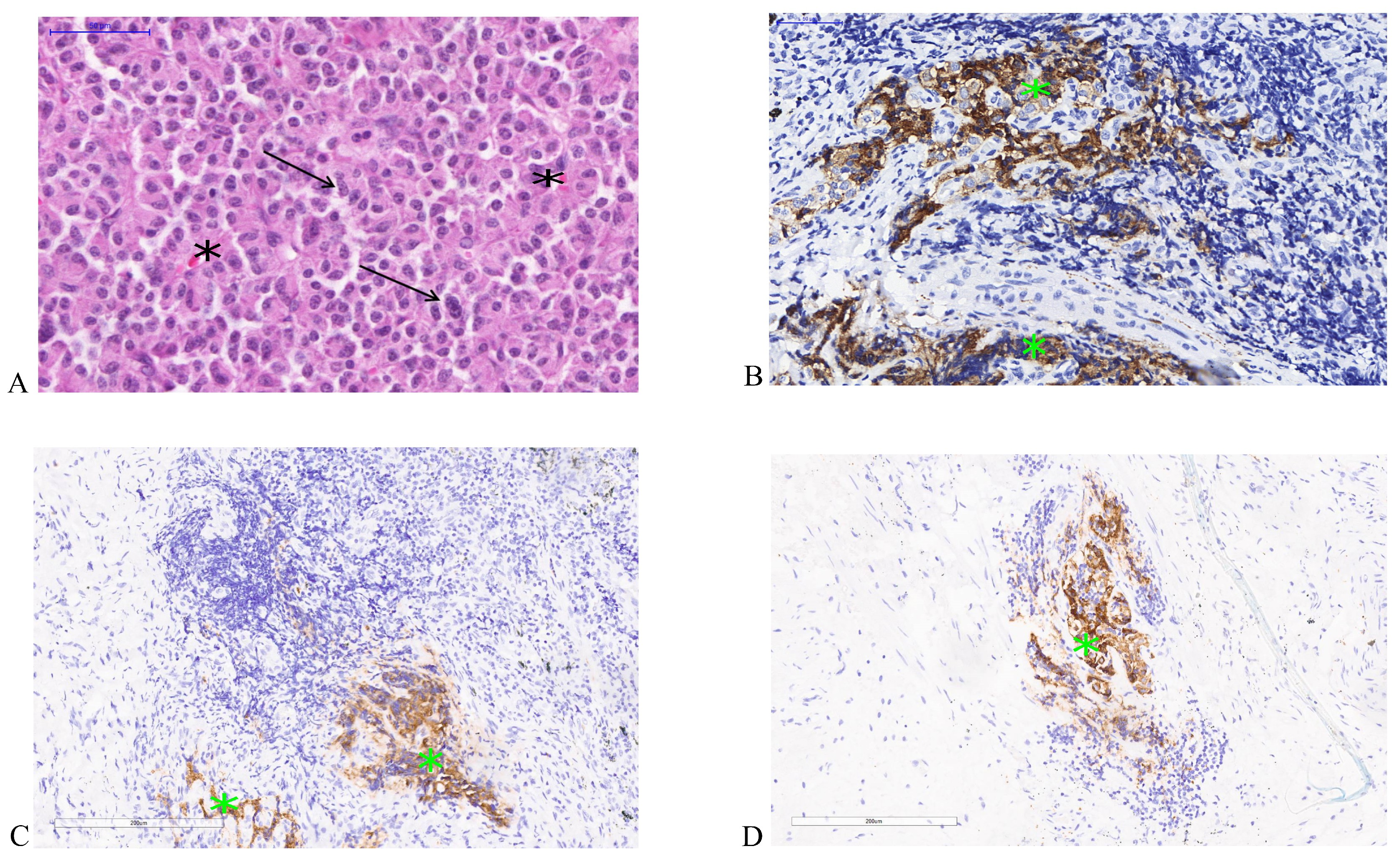
Fig. 1. (A) Haematoxylin and Eosin (H&E) stained section of the low grade, typical carcinoid tumour of the lung. Chromatin in cell nuclei show evident "salt and pepper" patterning as indicated by black arrows. Tumour shows characteristic organoid tumour growth with vascularization (indicated by black stars *) and absence of necrosis and lack of mitotic activity, 400X magnification. (B) Immunohistochemical stained section showing positivity for synaptophysin, a specific biomarker expressed in the typical subtype of carcinoid tumours, 400X magnification. (C) Immunohistochemical stained section showing positivity for chromogranin A, 200X magnification. (D) Immunohistochemical stained section showing positivity for cytokeratins (AE1/AE3), 200X magnification. The green stars* indicate positive immune staining areas.